The patient, 54-year-old Duong Thi Cuc of Lam Dong Province, suffered total necrosis of the femoral head at age three. She endured a lifelong leg-length discrepancy of 5–6 centimeters and progressively worsening pain and mobility. In recent years, the pain became debilitating, leaving her unable to walk and prompting her to seek treatment.
Clinical and imaging assessments confirmed advanced developmental dysplasia of the hip (DDH), a condition in which the acetabulum and femoral head fail to align anatomically, leading to partial or full dislocation, deformity, and chronic pain. Doctors determined the deformity had evolved to a severe stage, with nearly 50 years of structural changes rendering surgical reconstruction extraordinarily challenging.
Colonel Phan Dinh Mung, Director of the Orthopedic and Trauma Institute and Deputy Director of Military Hospital 175, said the decades-long deformity had reshaped the acetabular region. The bone was thin, soft, and structurally fragile, posing a high risk of fracture during surgery. Surrounding soft tissues and muscles were significantly contracted, shortening the left leg by up to 6 centimeters.

The femur presented equally formidable difficulties: a small femoral head and a narrow medullary canal meant standard prosthetic stems could not be used. Surgeons planned a corrective femoral osteotomy to reduce tension on the sciatic nerve while restoring limb length and hip function.
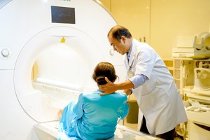
To tackle the complexities, the surgical team turned to 3D-printing — an increasingly important tool in orthopedic surgery. Using patient-specific imaging data, doctors printed a life-size resin model of the pelvis and proximal femur. The model allowed the team to determine the true acetabular geometry, estimate implant size, and identify the smallest viable stem compatible with the narrow canal.
“The 3D model enabled us to select the only prosthesis that would fit the femur and to design a plan to reconstruct the lost acetabular rim, a critical structure for bearing load after replacement,” Colonel Phan Dinh Mung said.

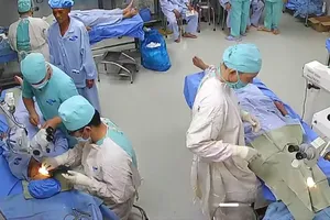
The operation lasted about 4.5 hours — shorter than predicted. Six days later, the patient showed no signs of sciatic nerve injury, the most feared complication.
Because the acetabulum was rebuilt with bone grafts, the patient will bear minimal weight for three to six months while the graft consolidates, before resuming normal movement on the artificial hip.